NEFRECTOMY
Nefrectomy
is surgical procedure where excision of the kidney is preformed. It is
indicated in the cases that defy surgical repair, such as:
·
Renal neoplasia,
·
Severe trauma (resulting in
uncontrollable bleeding or urine leaking),
·
Pyelonephritis (resistant to medical therapy),
·
Hydronephrosis,
·
Ureteral abnormalities (avulsion, stricture, rupture or
caliculi)
Before
nefrectomy, renal function in the opposite kidney should be assessed by
determining its glomerular filtration rate (GFR)*.
If
renal neoplasia is suspected, radiography (thoracic and abdominal) and
ultrasonography should be preformed to help rule out metastasis (including to
the opposite kidney). To avoid unintentional transaction, the opposite ureter
should always be identified. This is particularly critical when removing large
neoplasmas.
*Glomerular
filtration rate (GFR) is
the volume of fluid filtered from the renal (kidney) glomerular capillaries into the Bowman's capsule
per unit time.[1]
SURGICAL ANATOMY
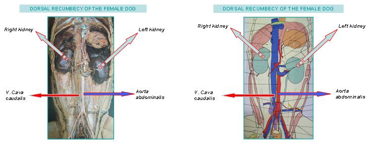
- The kidneys lies in
the retroperioneal space lateral to the aorta and caudal vena cava. They have
fibrouse capsule and are held in position by subperitoneal connective
tissue. The cranial pole of the
right kidney lies at the level of the 13th rib. In an average
sized dog, the cranial pole of the left kidney lies about
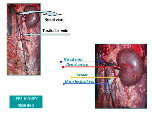
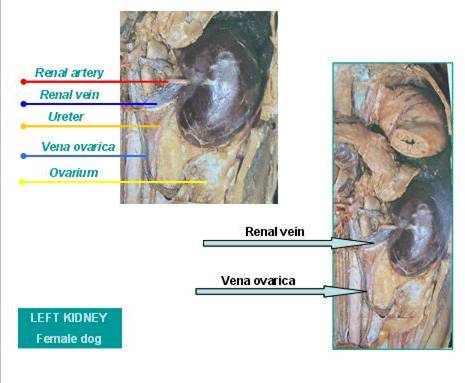
- The renal artery
normally bifurcates into dorsal and ventral branches but variations are
common
- The ureter begins at the renal pelvis and
enters the dorsal surface of the bladder.
NOTE: The anatomy of the renal blood vessels is highly variable, so care is
needed when ligate these vessels during nefrectomy.
The left ovarian and testicular veins drain into the renal vein so they
should not be ligated in intact dogs.
SURGICAL TECHNIQUES
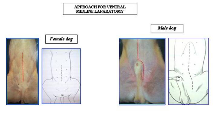
- Make ventral midline skin
incision begging from xyphoid and extending caudally towards the pubic
area. In male dogs extend it to prepucium and curve it to the left or
right side and extend to pubic area. After making the incision on the skin
blunt dissection is made to reach to the linea alba. The line is easier to find near the umbilicus
since it becomes thinner near the pubis. Then make incision with scalpel
on linea alba. Using the probe
and scalpel expand the incision to the pubis.
-
The right kidney can be exposed by elevating the duodenum and displacing the other loops of intestine on the animal’s left side. The left kidney can be exposed by elevating the mesocolon so that the small intestine is retracted to the animal’s right side.Female dog
- Grasp the peritoneum
over the kidney and incise it. Free the kidney from its sublumbar
attachments, using combination of blunt and sharp dissection. Elevate the
kidney and retract medially to locate the renal artery and vein on the
renal hilus. After identifying renal artery double ligate it with
absorbable or nonabsorbable suture close to abdominal aorta. Make sure
that all branches have been ligated. Identify the renal vein and ligate it
similarly. Left renal vein ligate above the drain of ovarian or testicular
vein. Ligate the ureter near the bladder with absorbable material. Use the
transfixation ligature to make sure it doesn’t slip off after cutting the vessels and
ureter.
- Close abdomen placing
interrupted sutures (X suture).
Incorporate full thickness bites of abdominal wall in the sutures
if the incision is made through linea alba. If the incision is lateral to
linea alba and muscular tissue is exposed (i.e. paramedian incision) close
the external rectus sheath without including muscle in sutures. Use the
absorbable sutures. Close subcutaneous tissue with absorbable suture
placing continuous sutures. For skin use nonabsorbable sutures placing
interrupted sutures.
NOTE: Sutures that are going to be used in
this surgery are explained and presented in ’’ SUTURE KNOTS AND PATTERNS ’’
REFERENCES:
- ^ Physiology at
MCG 7/7ch04/7ch04p11 - "Glomerular
Filtration Rate"
- Theresa Welch Fossum; Small Animal Surgery (second edition); 2002
Mosby;
- Stanley H.
Done, Peter C. Goody, Susana A. Evans, Neil C. Stickland; Color atlas of Veterinary;
volume 3; The dog and cat; 1996 Mosby, Missouri